Thoracoscopy
Thoracoscopy – A Minimally Invasive Procedure for Diagnosing and Treating Chest Diseases
Thoracoscopy, also known as Video-Assisted Thoracoscopic Surgery (VATS), is a minimally invasive medical procedure used to examine the pleural cavity (the space between the lungs and the chest wall). It allows doctors to visualize the lungs, pleura, diaphragm, and mediastinum using a small camera and specialized instruments inserted through tiny incisions in the chest wall. Thoracoscopy plays a vital role in both the diagnosis and treatment of various thoracic conditions.
Thoracoscopy is often performed when less invasive methods—like imaging or needle biopsy—do not provide sufficient information. One of its most common uses is to investigate unexplained pleural effusion (fluid buildup around the lungs). With direct visualization, doctors can inspect the pleura for signs of infection, inflammation, tuberculosis, or cancer, and take targeted biopsies to obtain accurate tissue samples. This greatly improves diagnostic accuracy, particularly in diseases like malignant pleural mesothelioma, metastatic cancers, or chronic infections.
In addition to diagnosis, thoracoscopy is used for several therapeutic interventions. These include drainage of pleural fluid, removal of pleural adhesions, pleurodesis (a procedure to prevent recurrent fluid buildup in the pleural space), and even wedge resections of lung tissue for suspected early-stage cancer. Because thoracoscopy avoids the need for large incisions, it results in less postoperative pain, quicker recovery, and shorter hospital stays compared to traditional open surgery (thoracotomy).
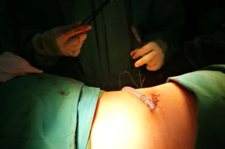
Thoracoscopy is typically performed under general anesthesia in an operating room, although in select cases, it can be done under local anesthesia with sedation, especially for diagnostic procedures. Patients are usually positioned on their side, and a small camera (thoracoscope) is inserted into the chest cavity, allowing the surgeon or pulmonologist to view high-resolution images on a monitor.
While generally safe, thoracoscopy does carry some risks, including bleeding, infection, air leaks, and in rare cases, injury to nearby organs. However, complications are relatively uncommon when performed by experienced professionals.
In summary, thoracoscopy is an invaluable technique in thoracic medicine. It combines the benefits of direct visual assessment and tissue sampling with the advantages of a minimally invasive approach. As a result, thoracoscopy has become a gold standard for diagnosing and managing pleural and lung diseases with greater precision and reduced patient discomfort.